Gallbladder and Extrahepatic Bile Ducts - Overview

1. Gallbladder and Bile Duct Anatomy

Gallbladder Anatomy
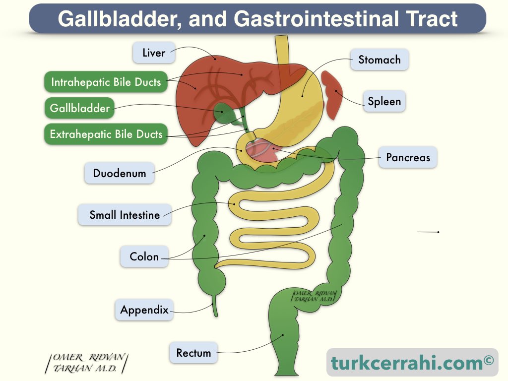
The gallbladder is a pear-shaped, 7-10 cm long, 30-50 ml capacity sac. The gallbladder is attached to the anterior and inferior surfaces of the liver. The gallbladder is histologically different from other gastrointestinal organs; it lacks the muscularis mucosa and submucosa. The gallbladder stores bile produced by the liver and concentrates it by absorbing water. The gallbladder contracts and empties the concentrated bile into the duodenum when food is consumed. Despite its name, the gallbladder does not produce bile itself.
Bile Duct Anatomy
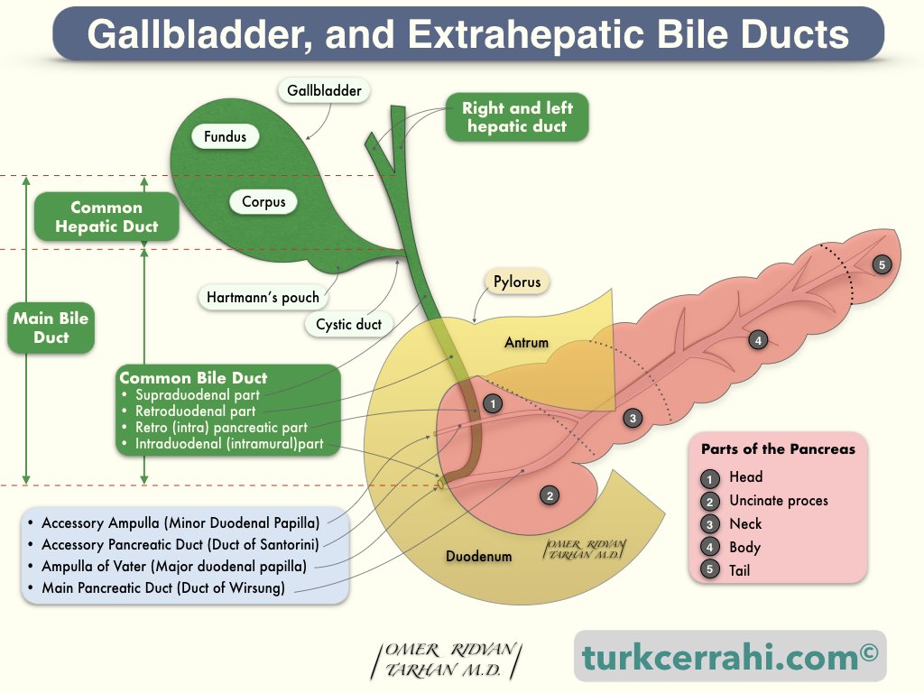
Extrahepatic bile ducts consist of the right and left hepatic ducts, the main hepatic duct, the cystic duct, and the common bile duct. The hepatic duct is 1–4 cm long. The cystic duct is joined to the hepatic duct to form the common bile duct. The common duct joins the main pancreatic duct (Wirsung) before opening into the duodenum. This biliopancreatic common channel runs inside the duodenal wall for 1 to 2 cm. The biliopancreatic common channel is wider just before it opens the duodenum (Ampulla Vater). Here, the mucosa of the duodenum is raised (Papilla Vater, duodenal papilla).
2. Gallbladder Disease
Gallstones (cholelithiasis) are the most common gallbladder disease. Gallstones can cause abdominal pain and acute cholecystitis (inflammation of the gallbladder).
Acute cholecystitis can be treated either with antibiotics or surgery. Acute cholecystitis can result in gallbladder gangrene, empyema (a pus-filled gallbladder), and perforation.
Gallstones, if they fall into the biliary tract, cause obstructive jaundice and biliary pancreatitis.
Although rare, gallbladder inflammation without gallstones may occur and is called acalculous cholecystitis. Acalculous cholecystitis is common in the elderly and debilitated patients. It is extremely dangerous and easily perforates.
Rarely, the gallbladder does not work properly, causing abdominal pain (biliary dyskinesia).
Gallbladder polyps are usually detected incidentally during ultrasonography. Some authors recommend cholecystectomy for gallbladder polyps larger than 7 mm, while others recommend 10 mm.
3. Biliary Tract Disease
Bile duct stones (choledocholithiasis) are the most common biliary tract diseases. The origin of a biliary stone is almost always a stone that falls out of the gallbladder, called the secondary bile duct stone. Bile duct stones rarely develop inside the biliary tract and are called primary bile duct stones. Bile duct stones (choledocholithiasis) can cause jaundice (due to bile duct obstruction), cholangitis (biliary tract inflammation), and pancreatitis (a stone sitting on the bulb).
The most common cause of bile duct strictures today is gallbladder and biliary tract surgery.
Primary sclerosing cholangitis is characterized by multiple intrahepatic and/or extrahepatic bile duct strictures. Normal or slightly dilated segments are also observed. Primary sclerosing cholangitis is an autoimmune disease (the body attacks its own tissues).
Extrahepatic Bile Duct Cancers
Extrahepatic bile duct cancers are traditionally classified into three types:
- Gallbladder cancer
- Extrahepatic biliary tract cancer
- Ampulla of Vater cancer.
Gallbladder cancers are rare but deadly cancers. Approximately half of gallbladder cancers can still be detected during cholecystectomy surgery despite the use of advanced imaging techniques. 5–10% of biliary tract cancers (cholangiocarcinomas) arise from intrahepatic bile ducts, while 90–95% arise from extrahepatic bile ducts. The most typical finding of bile duct cancer is painless jaundice.